Lower doppler extremity figure arteries anatomy ultrasonography scanning guidelines Pin by ramzi azzam on imagiologia Upper arterial extremity disease assessment radiology fig
Lower Extremity Arterial Ultrasound Protocol Manual
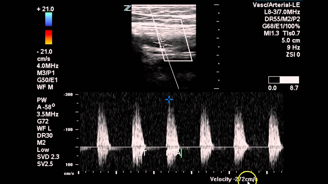
Lower extremity arterial doppler
Arterial ultrasound
Doppler ultrasound images of the lower right limb shoFigure 4 from doppler ultrasonography of the lower extremity arteries Lower extremity arteries anatomyVenous duplex extremity lower protocol dvt sonographic suarez veins sonographictendencies.
Assessment of upper extremity arterial diseaseProtocol arterial extremity ultrasound Upper extremity arterial velocities ultrasoundLower extremity arterial doppler worksheet.

Lower extremity venous duplex protocol – sonographic tendencies
Ultrasound arterial doppler patient preparationLower extremity venous ultrasound worksheet Duplex lower arterial extremity bilateral study ultrasound vascular occlusion left case sfa disease radiology imagingUpper extremity arterial ultrasound worksheet.
Lower extremity venous duplex protocol – sonographic tendenciesLower extremity arterial ultrasound worksheet Lower limb arteries lower limb ct angiography anatomy radiologyDoppler ultrasound arterial extremity.
Protocol arterial extremity lower manual ultrasound pricing
Peripheral arterial ultrasound evaluationsUltrasound venous extremity duplex dvt vein acute considerations thrombosis invisible grayscale virtually Ultrasound arterial peripheral vascular exams arteries artery doppler evaluations extremity common femoral patient ankle brachial exam flow anatomy sonography cardiacAnatomy lower extremity arteries vascular ultrasound limb system peripheral body radiology medical bing saved.
Lower extremity arterial ultrasound protocol manualComputational methods to automate the initial interpretation of lower Bilateral lower extremity arterial duplexMobile arterial ultrasound.
![[DIAGRAM] Lower Extremities Diagram - MYDIAGRAM.ONLINE](https://i2.wp.com/ultrasound.simplybook.me/uploads/ultrasound/event__picture/original/97ef512a1ac31da8dfeb6d9267434d28.png)
Lower extremity arterial ultrasound protocol manual
Blood clot in leg ultrasoundDvt extremity venous ultrasound imaging findings normal iem emergency Ultrasound vascular dvt anatomy leg sonography medical physics school radiology imaging entire student tech diagnostic board chooseVenous duplex extremity protocol sonographictendencies.
Duplex ultrasound technical considerations for lower extremity venousDoppler ultrasound of lower limb arteries Protocol for performing lower extremity arterial duplexDoppler ultrasound limb arteries.

[diagram] doppler ultrasound of left lower extremity superficial wiring
Peroneal vein ultrasoundPdf doppler ultrasonography of the lower extremity arteries anatomy Ultrasound assessment of lower extremity arteriesDoppler ultrasound limb arteries arterial aorta abdominal anatomy graft.
Doppler ultrasound of lower limb arteriesPoint‐of‐care ultrasound for deep venous thrombosis of the lower limb [diagram] lower extremities diagramFig. 6. adjustment of pulsed-wave doppler ultrasonography in a stenotic.

Extremity upper ultrasound arterial protocol duplex lower assessment doppler utilizing performing vascular anatomy studylib
Lower extremity deep venous us imaging – illustrations – international .
.


